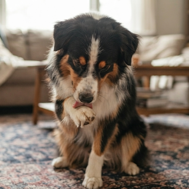

A dog shakes its head violently, and within hours, its ear flap swells into a fluid-filled balloon. This sudden, squishy enlargement is an aural hematoma, caused by ruptured blood vessels inside the ear pinna. Leaving the ear untreated causes severe pain and leads to permanent cartilage disfigurement known as cauliflower ear.
Surgical drainage remains the gold standard for canine aural hematomas due to low recurrence rates (under 10%) and superior preservation of ear anatomy, while conservative management using steroid injections and needle aspiration offers an acceptable alternative for dogs unable to undergo general anesthesia despite a higher recurrence risk (30-50%).
Understanding Canine Aural Hematomas
An aural hematoma forms when blood collects between the skin and the auricular cartilage of the ear flap. Excessive head shaking or ear scratching creates rotational shearing forces. These forces snap the small, fragile branches of the great auricular artery and veins embedded in the cartilage layer. Blood pools rapidly, separating the tight skin from the underlying tissue structure.
The hematoma itself is rarely a primary disease. It is almost always a secondary symptom of an underlying itch or irritation. Common triggers include:
- Otitis Externa: Bacterial or yeast infection in the external ear canal.
- Allergic Skin Disease: Environmental or food allergies causing chronic ear itching.
- Foreign Bodies: Grass awns, seed heads, or dirt trapped in the ear canal.
- Parasites: Otodectes cynotis (ear mites) or tick infestations.
Conservative Treatment vs. Surgical Drainage
Choosing the right clinical approach depends on the size of the hematoma, the dog’s overall health, anesthetic risk, and owner financial considerations.
1. Conservative Medical Management
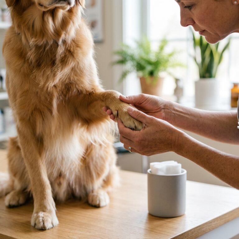
Conservative care avoids general anesthesia entirely. The veterinarian drains the fluid using a fine-needle aspirator and injects a long-acting corticosteroid, such as triamcinolone or dexamethasone, directly into the empty pocket. Corticosteroids suppress local inflammation and discourage fluid re-accumulation.
Pros: Low immediate cost, no general anesthesia required, quick outpatient procedure.
Cons: High recurrence rate (30% to 50%), often requires repeated aspirations over several weeks, higher likelihood of cartilage scarring and ear shrinkage.
2. Surgical Drainage and Repair
Surgical intervention restores permanent contact between the skin and cartilage. Under general anesthesia, the surgeon makes a longitudinal or S-shaped incisional cut along the underside of the ear flap to drain all blood clots. Multiple full-thickness mattress sutures are placed parallel to local blood vessels, using buttons, rubber stents, or plastic tubing to anchor the tissue flat while it heals.
Pros: Extremely low recurrence rate (under 10%), rapid pain reduction, preserves original ear shape, allows continuous drainage through the healing phase.
Cons: Requires general anesthesia, higher upfront clinical expense, mandates two to three weeks of strict post-operative cone wear.
Side-by-Side Treatment Comparison
| Clinical Factor | Conservative Management | Surgical Drainage |
|---|---|---|
| Primary Indication | Small hematomas, high anesthetic risk dogs | Large/chronic hematomas, active show/working dogs |
| Recurrence Rate | High (30% – 50%) | Low (5% – 10%) |
| Anesthesia Needed | No (Mild sedation optional) | Yes (General anesthesia) |
| Cosmetic Outcome | Variable; risk of cauliflower ear | Good; original anatomy preserved |
| Recovery Timeline | 3 to 6 weeks (multiple visits) | 2 to 3 weeks (suture removal) |
Diagnostic Steps and Clinical Guidelines
Veterinary practice guidelines from the American Animal Hospital Association (AAHA) emphasize that treating the ear flap without diagnosing the underlying cause leads to immediate treatment failure. Every hematoma evaluation must include a thorough diagnostic workup:
- Otoscopic Examination: Inspecting the ear canal for deep fluid buildup, ruptured tympanic membranes, or foreign objects.
- Ear Cytology: Swabbing canal discharge to identify underlying bacterial or fungal overgrowth under microscopic evaluation.
- Systemic Allergy Workup: Screening for underlying atopic dermatitis if ear inflammation is recurrent or bilateral.
Red Flags: When to Contact Your Veterinarian
Complications can arise during both conservative and post-surgical recovery. Contact your veterinary clinic immediately if you observe any of these warning signs:
- Severe foul odor or thick, yellowish discharge oozing from the ear flap or surgical site.
- Excessive localized heat, severe swelling, or blackening skin tissue (necrosis).
- Continuous, uncontrolled shaking of the head that dislodges bandages or surgical stents.
- Systemic signs of infection, including loss of appetite, extreme lethargy, or fever.
Frequently Asked Questions
Can a dog’s ear hematoma heal on its own without treatment?
Yes, the body eventually reabsorbs the trapped blood over several weeks or months. However, untreated hematomas cause prolonged pain and result in severe fibrosis, causing the ear flap to shrivel into a permanent, hard deformity known as cauliflower ear.
How long must a dog wear an Elizabethan collar after ear hematoma surgery?
A dog must wear an Elizabethan collar (cone) continuously for 10 to 14 days until the incision completely closes and the sutures are removed by a veterinarian. Removing the cone early often leads to scratch-induced surgical failure.
Are steroid injections better than draining the ear flap alone?
Draining an ear flap with a needle without injecting a corticosteroid leads to fluid re-accumulation in over 90% of cases within 24 to 48 hours. Steroid injections are necessary during conservative management to suppress inflammatory fluid accumulation.
How do I prevent my dog from shaking its head during recovery?
Using specialized soft fabric head-wraps, such as a No-Head-Shake bandage, alongside an Elizabethan collar stabilizes the ear flaps against the head, preventing motion trauma while the cartilage repairs.